From Bicycles to CBCT
Boys and girls love their toys, and I am no exception. Christmas of 1952 was unforgettable when a brand new Rollfast bicycle appeared next to our Christmas tree. My freedom flourished as I rode faster than I could ever walk or run.
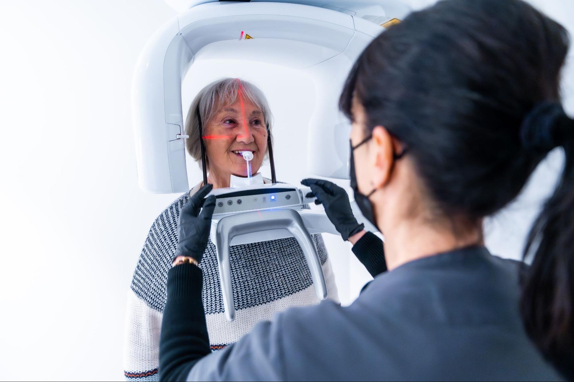
Decades later, I felt the same excitement when our office in Westlake Village, CA became an early adopter of CBCT technology. I called the machine my “Ferrari,” and one of my patients even gave me a Ferrari sticker to place on it.
The Freedom of Seeing Clearly
Just as a bicycle gave me the freedom to explore as a child, CBCT gave me the freedom to see everything in detail on an x-ray. It brought a smile to my face and opened opportunities to understand what had previously been invisible.
Gone are the days of speculating about airway size or shape. With CBCT we can now see:
All of this is available in three dimensions, without anything invasive.
A Leap Beyond Earlier Technology
In the 1990s, I used a Quint Sectograph to capture TMJ images for my patients. It dramatically increased diagnostic capability at the time.
But CBCT eclipsed that technology. It provided massively more information about condylar position, disc placement, and condylar health. With CBCT, we can slice and analyze images in ways never before possible. We can assess airways in multiple cross-sections and visualize tooth positions in all three planes of space.
More Than a Machine
Like every new technology, CBCT comes with a learning curve. We must train our eyes to interpret what we see, distinguish between normal and abnormal, and most importantly, understand what the findings mean for the patient.
CBCT is not just about owning a Ferrari. It is about having the skill to drive it. When used with trained expertise, CBCT elevates diagnosis and treatment planning—and everyone benefits.